
Glaucoma is one of the leading causes of blindness and visual impairment in the US, and can affect patients of all ages. Many glaucoma patients do not experience any symptoms and may not be aware that they have the disease. Glaucoma actually refers to a group of diseases that cause damage to the optic nerve as a result of increased pressure within the eye. Occasionally it can also be caused by injury, blocked blood vessels or inflammatory conditions of the eye.
There are two main types of glaucoma, open-angle and angle-closure. Open-angle glaucoma is the most common type of glaucoma and involves fluid in the eye not draining properly through the trabecular meshwork. Angle-closure glaucoma involves a sudden buildup of pressure in the eye and poor drainage because the angle between the iris and the cornea is too narrow. This less common type of glaucoma can cause eye pain, redness and halos around lights.
Many patients do not experience any symptoms during the early stages of open angle glaucoma. This makes it difficult for many patients to know if they have the disease. As glaucoma progresses, patients may experience a loss of peripheral or side vision. Our goal is to diagnose and treat glaucoma before vision is lost.
Diagnosing Glaucoma
While some patients may experience symptoms from glaucoma as the disease progresses, others do not learn they have the condition until they undergo a routine eye exam. There are several different exams performed to diagnose glaucoma, including a visual field test and optic nerve measurements. Other tests will also be performed, such as tonometry to measure the pressure inside the eye and pachymetry to measure the thickness of the cornea.
Treatment for Glaucoma
Glaucoma Rx Eye Drops
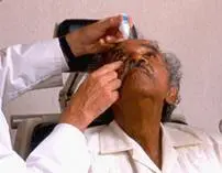
Eye drops are used to reduce fluid production in the front of the eye or to help drain excess fluid. Eye drops are usually well tolerated, but can cause temporary stinging or blurred vision. Patients should tell their doctor about any allergies or illnesses they have to minimize the risk of side effects.
Once glaucoma has been diagnosed, treatment should begin as soon as
possible to help minimize the risk of permanent vision loss. There is no cure for glaucoma, so treatment focuses on preventing further damage from occurring. Most cases of glaucoma can be treated with eye drops, laser surgery or microsurgery. The best treatment for your individual case depends on the type and severity of the disease, and can be discussed with your doctor.
Glaucoma Rx Eye Drops
Eye drops are used to reduce fluid production in the front of the eye or to help drain excess fluid. Eye drops are usually well tolerated, but can cause temporary stinging or blurred vision. Patients should tell their doctor about any allergies or illnesses they have to minimize the risk of side effects.
Laser Surgery
Laser surgery for glaucoma aims to increase the outflow of fluid from the eye or eliminate fluid blockages through laser selective laser trabeculoplasty, iridotomy or cyclophotocoagulation. Selective laser trabeculoplasty (SLT) is an in-office procedure that reduces intraocular pressure in patients with glaucoma. The laser is applied through a special contact lens to the drainage system of the eye where it stimulates a biochemical change that improves the outflow of fluid from the eye.
MIGS has become a commonly used abbreviation in the glaucoma world. It stands for minimally invasive glaucoma surgery.
MIGS
The goal of all glaucoma surgery is to lower eye pressure to prevent or reduce damage to the optic nerve.
Why MIGS?
Standard glaucoma surgeries — trabeculectomy and ExPRESS shunts, external tube-shunts like the Ahmed and Baerveldt styles — are major surgeries. While they are very often effective at lowering eye pressure and preventing progression of glaucoma, they have a long list of potential complications.
The MIGS group of operations have been developed in recent years to reduce some of the complications of most standard glaucoma surgeries.
MIGS procedures work by using microscopic-sized equipment and tiny incisions. While they reduce the incidence of complications, some degree of effectiveness is also traded for the increased safety.
The MIGS group of operations are divided into several categories:
- Miniaturized versions of trabeculectomy
- Trabecular bypass operations
- Totally internal or suprachoroidal shunts
- Milder, gentler versions of laser photocoagulation
Microtrabeculectomies
Using tiny, microscopic-sized tubes that can be inserted into the eye and drain fluid from inside the eye to underneath the outer membrane of the eye (conjunctiva), two new devices seem to make the trabeculectomy operation safer. These devices (the Xen Gel Stent and PRESERFLO [formerly InnFocus] Microshunt) have shown excellent pressure lowering with improved safety over trabeculectomy in studies done outside the United States. (2019 update: These devices have recently been approved for use in the US).
Trabecular Surgery
Most of the restriction to fluid drainage from the eye rests in the trabecular meshwork. Several operations have been devised using tiny equipment and devices to cut through the trabecular meshwork without damaging any other tissues in the ocular drainage pathway. Using a special contact lens on the eye, a tiny device is inserted into the eye through a tiny incision into the trabecular meshwork under high power microscopic control. The trabecular meshwork can either be destroyed (Trabectome or Trab360) or bypassed using a tiny snorkel-like device (the iStent). These procedures are FDA-approved but generally don’t get the eye pressure very low so are most useful in early to moderate stages of glaucoma.
Suprachoroidal Shunts
Using tiny tubes with very small internal openings, the front of the eye is connected to the suprachoroidal space between the retina and the wall of the eye (Glaukos shunts) to augment the drainage of fluid from the eye. This operation has relatively few serious complications and lowers pressures enough to be useful even in moderately severe glaucoma.
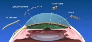
Placement of some MIGS devices
New Laser Procedures
Previously, laser cyclophotocoagulation was reserved for advanced glaucoma that could not be controlled despite trabeculectomy or tube shunts. The procedures were designed to reduce the fluid-forming capacity of the eye by targeting the delicate tissue (ciliary body) that makes the fluid. They sometimes produced severe inflammation that could reduce vision. Two recent additions to the laser treatment procedures have proven useful even before the glaucoma is far advanced. These are endocyclophotocoagulation and micropulse cyclophotocoagulation. These procedures may be discussed in a future article.
Summary
Several new approaches to glaucoma surgery show promise for better safety. As with all new procedures, time and much follow-up study are required to see which ones will remain useful for helping glaucoma patients long-term.
Glaucoma Surgeries
Glaucoma surgeries include the delicate, microscopic incisional trabeculectomy (with or without ExPRESS microshunt implantation), tube shunt implantation (a shunt is a glaucoma drainage device), and cyclophotocoagulation . There are also newer procedures called MIGS, or minimally invasive glaucoma surgery . Each type of glaucoma surgery has its unique uses, advantages and disadvantages.When considering glaucoma surgery, you should have a detailed discussion with your surgeon about the risks and benefits of the specific surgery you may undergo.
When considering glaucoma surgery, you should have a detailed discussion with your surgeon about the risks and benefits of the specific surgery you may undergo. In this article, we will discuss the risks and benefits of glaucoma surgery in general.
Preventing Glaucoma
While there are no known ways to prevent glaucoma from developing, regular screenings and early detection are the best forms of protection against the harmful damage that the disease can cause. While anyone can develop glaucoma, some people are at a higher risk for developing disease. These people may include those who:
- Are over the age of 60
- Are African American or Hispanic over the age of 40
- Have a family history of glaucoma
- Have diabetes
Patients should have a comprehensive dilated eye exam at least once every two years, especially if they have a higher risk of developing glaucoma. Older patients may be encouraged to be tested more frequently.